Retinal Vessel Narrowing: What It Means and When to Seek Help
Being told that blood vessels at the back of your eyes appear narrowed can sound alarming. Online advertisements make the finding even more frightening by claiming that the vessels will continue shrinking until the eyes are “starved” and vision disappears.
That is not an accurate explanation.
Retinal vessel narrowing can be associated with high blood pressure, aging, diabetes, inflammation, and other vascular factors. Its significance depends on which vessels are affected, what other retinal changes are present, and the person’s overall health.
It is not a diagnosis of inevitable blindness, and it does not prove that floaters or wavy vision are caused by narrowed vessels.
Those symptoms have several possible causes, some of which require urgent treatment. A supposed “eye-restoring ritual” cannot safely diagnose or treat them.
What are retinal blood vessels?
The retina is the light-sensitive layer of tissue at the back of the eye. It converts light into electrical signals that travel to the brain through the optic nerve.
Like other tissues, the retina depends on a network of arteries, veins, and tiny capillaries.
- Retinal arteries carry oxygenated blood toward retinal tissue.
- Retinal veins carry blood away from the retina.
- Capillaries allow oxygen and nutrients to reach retinal cells.
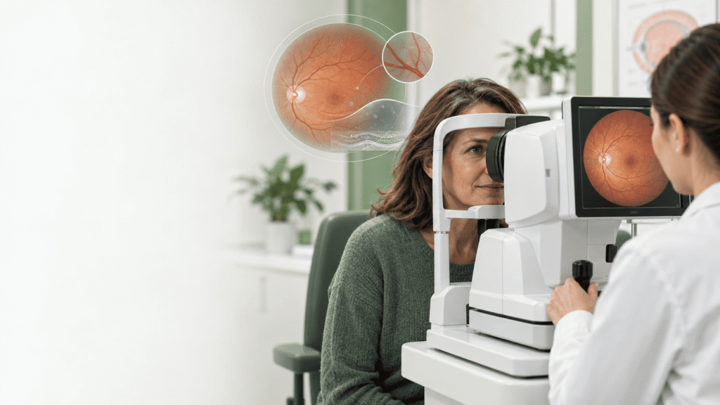
An eye doctor can examine parts of this network through the pupil. Because retinal vessels can be observed without surgery, an eye examination may reveal clues about both eye health and systemic vascular health.
What does “vessel narrowing” mean during an eye exam?
The phrase commonly refers to retinal arteriolar narrowing—the appearance of relatively narrow small arteries in the retina.
The finding should be interpreted in context. It may be:
- A long-standing anatomical characteristic
- Associated with aging
- Related to chronic high blood pressure
- Accompanied by other signs of hypertensive retinopathy
- Part of a broader retinal or vascular condition
- A measurement generated by retinal photography or imaging software
“Narrowing” does not necessarily mean that a vessel is steadily closing until no blood can pass through it. Nor does the phrase alone predict that someone will lose vision within five years.
The relevant questions are:
- Which vessels appear narrowed?
- Is the finding present in one or both eyes?
- Are there haemorrhages, swelling, leakage, deposits, or abnormal vessel crossings?
- Is the optic nerve affected?
- Does the person have high blood pressure, diabetes, high cholesterol, or cardiovascular disease?
- Has the appearance changed over time?
- Are there symptoms?
A proper interpretation requires the complete examination, not one phrase taken from a report.
Does retinal vessel narrowing cause floaters?
Usually, floaters have a different explanation.
Floaters are small spots, strands, circles, cobwebs, or shadows that move across the field of vision. They often occur because strands within the vitreous—the gel-like substance inside the eye—cast shadows on the retina.
Vitreous changes become more common with age. A posterior vitreous detachment can produce new floaters and flashes as the vitreous separates from the retina.
The National Eye Institute explains that vitreous detachment commonly causes a sudden increase in floaters and may also cause flashes of light in peripheral vision. National Eye Institute
Floaters may also be associated with:
- Retinal tears
- Retinal detachment
- Bleeding inside the eye
- Eye inflammation
- Diabetic retinopathy
- Eye injury
- Previous surgery
Retinal vessel narrowing is not a universal explanation for floaters.
When are floaters an emergency?
Long-standing floaters that have already been examined may be harmless. A sudden change is different.
Seek urgent eye care if you experience:
- Many new floaters appearing suddenly
- Flashes of light
- A dark curtain or shadow across your vision
- A new blurry area
- Sudden loss of peripheral vision
- Vision loss after an eye injury
These symptoms may indicate a retinal tear or detachment.
A retinal detachment happens when the retina separates from its normal position. It can permanently damage vision if treatment is delayed. The National Eye Institute identifies sudden floaters, flashes, and a curtain-like shadow as major warning signs. National Eye Institute retinal-detachment guidance
Do not wait for a supplement, exercise, or online ritual to make these symptoms disappear.
Why can straight lines look wavy?
When straight lines appear bent, distorted, rippled, or broken, the symptom is called metamorphopsia. It usually points toward a problem affecting the macula—the central part of the retina responsible for detailed vision.
Possible causes include:
- Age-related macular degeneration
- Macular oedema
- Macular pucker
- Macular hole
- Diabetic retinal changes
- Retinal-vein occlusion
- Inflammation
- Certain inherited or structural retinal disorders
The National Eye Institute identifies wavy or crooked lines as a warning sign of late age-related macular degeneration and recommends prompt eye evaluation. National Eye Institute AMD guidance
A macular pucker can also make text, objects, and straight lines appear distorted. National Eye Institute
Wavy vision should not be assumed to come from blood vessels “shrinking.” It requires examination because treatment depends on the underlying condition.
What is hypertensive retinopathy?
High blood pressure can affect small blood vessels throughout the body, including those in the retina.
Possible retinal findings associated with hypertension include:
- Generalised or focal narrowing of retinal arteries
- Changes where arteries cross over veins
- Retinal haemorrhages
- Cotton-wool spots
- Lipid deposits
- Optic-disc swelling in severe cases
Mild vessel changes may not cause noticeable symptoms. More severe findings can indicate significant vascular injury and require prompt evaluation of blood pressure and cardiovascular health.
An eye doctor may advise the patient to consult a primary-care doctor or cardiovascular specialist. The goal is not merely to treat the eye but to control the condition affecting the blood vessels.
Is retinal vessel narrowing a stroke warning?
Retinal vascular findings can be associated with systemic vascular risk, but they are not a simple countdown to stroke or blindness.
High blood pressure, diabetes, smoking, high cholesterol, atrial fibrillation, and other vascular conditions can raise the risk of both retinal disease and stroke. Finding retinal changes may therefore prompt a review of these risk factors.
That does not mean everyone with relatively narrow retinal vessels is about to have a stroke.
Emergency assessment is required for symptoms such as:
- Sudden weakness or numbness on one side
- Facial drooping
- Difficulty speaking or understanding speech
- Sudden severe imbalance
- Sudden vision loss
- Double vision
- A severe, unexplained headache
- Confusion or loss of consciousness
Sudden painless loss of vision in one eye can also be an ocular emergency and should not be monitored at home.
What is retinal vein occlusion?
Retinal vein occlusion occurs when a vein carrying blood away from the retina becomes blocked. It is different from general retinal vessel narrowing.
Central retinal vein occlusion affects the main retinal vein, while branch retinal vein occlusion affects one of its branches.
Symptoms may include:
- Sudden blurry vision
- Partial vision loss
- Distortion
- A dark or missing area
- Symptoms in one eye
The National Eye Institute states that central retinal vein occlusion is diagnosed through a dilated eye examination and imaging. Treatment may include eye injections or laser treatment, depending on the complications. National Eye Institute
No home ritual can physically unblock every retinal vessel or reverse all resulting damage.
Can ordinary eye doctors see retinal vessels?
Yes. The claim that most optometrists cannot see retinal vessels during a regular examination is misleading.
Eye-care professionals can examine the retina using tools such as:
- Direct or indirect ophthalmoscopy
- Slit-lamp examination with specialized lenses
- Dilated fundus examination
- Retinal photography
- Optical coherence tomography
- OCT angiography
- Fluorescein angiography in selected cases
Not every patient requires every test.
A dilated examination allows a wider view of the retina. Retinal photography documents visible structures for comparison. Optical coherence tomography provides cross-sectional images of retinal layers. Angiography may help evaluate blood flow, leakage, or abnormal vessels.
The clinician selects tests based on the symptoms, medical history, and initial findings.
Why might an eye doctor recommend monitoring?
Monitoring does not necessarily mean that the doctor is ignoring a dangerous problem.
Some retinal findings are stable, mild, or unlikely to benefit from immediate intervention. In those situations, scheduled observation may be safer than unnecessary treatment.
Monitoring may involve:
- Repeat dilated examinations
- Comparing retinal photographs
- Measuring vision
- Checking eye pressure
- Repeating OCT imaging
- Monitoring blood pressure or blood sugar
- Watching for new symptoms
However, the patient should understand the working diagnosis, expected course, follow-up interval, and warning signs that require earlier reassessment.
If the explanation is unclear, ask:
- What exactly did you observe?
- What is the likely cause?
- Is it affecting my vision?
- Do I need medical tests or a blood-pressure check?
- What symptoms should make me contact you immediately?
- Would a retinal specialist’s opinion be useful?
Seeking a second professional opinion is reasonable when a serious diagnosis remains unclear. Replacing professional care with an online sales video is not.
Can a natural ritual widen retinal vessels?
There is no single home ritual proven to widen retinal vessels and restore all forms of vision loss.
Vision changes can result from damage to the cornea, lens, optic nerve, vitreous, macula, retina, blood vessels, or visual pathways in the brain. One remedy cannot correct all these structures.
A person who reports that floaters and distortion disappeared after a few days does not prove that retinal blood vessels changed. Symptoms can fluctuate, and testimonials cannot establish cause and effect.
Treatment may involve:
- Managing blood pressure
- Controlling blood glucose
- Treating abnormal cholesterol
- Smoking cessation
- Anti-VEGF eye injections
- Steroid treatment
- Laser treatment
- Retinal surgery
- Cataract surgery
- Corrective lenses
- Monitoring when treatment is unnecessary
- Treatment of an underlying inflammatory or neurological condition
The correct intervention depends on the diagnosis.
Can eye injections improve retinal disease?
For some retinal conditions, yes.
Anti-VEGF medicines can reduce abnormal blood-vessel growth, leakage, and retinal swelling. They may be used for wet age-related macular degeneration, diabetic macular oedema, and macular oedema related to retinal-vein occlusion.
The National Eye Institute explains that these injections block vascular endothelial growth factor, a protein that can contribute to abnormal vessels and leakage. National Eye Institute
Not every retinal finding requires injections, and only an ophthalmologist can determine whether they are appropriate.
How can you protect your vision after 40?
Arrange regular comprehensive eye examinations
Eye diseases such as glaucoma, diabetic retinopathy, and early macular degeneration may not cause obvious symptoms initially.
Ask your eye-care professional how frequently you should be examined based on your age, family history, health conditions, prescription, and previous findings.
Manage blood pressure
High blood pressure can damage blood vessels in the eyes and elsewhere. Follow your clinician’s recommendations for monitoring, medication, activity, nutrition, and follow-up.
Control diabetes
Elevated blood glucose can damage retinal vessels. People with diabetes should follow the eye-examination schedule recommended by their healthcare team.
Avoid smoking
Smoking increases the risk of several eye diseases, including age-related macular degeneration.
Protect your eyes from ultraviolet exposure
Use sunglasses that provide appropriate UV protection and wear a hat in strong sunlight.
Exercise and eat a balanced diet
Regular physical activity and a diet containing vegetables, fruit, protein, whole grains, and suitable healthy fats can support cardiovascular and general health. No single food or supplement guarantees protection from vision loss.
Know your family history
Tell your eye doctor about glaucoma, macular degeneration, retinal detachment, and inherited eye conditions in close relatives.
The bottom line
Retinal vessel narrowing deserves proper interpretation, but it does not mean that blindness is inevitable within five years.
Floaters, wavy lines, and blurry vision can arise from several different eye conditions. A sudden increase in floaters, flashes, a curtain-like shadow, or sudden vision loss requires urgent evaluation. New wavy vision also warrants prompt professional assessment.
Eye doctors can examine retinal vessels through dilated examinations and imaging. When they recommend monitoring, ask for a clear diagnosis, follow-up plan, and emergency warning signs.
Do not rely on an anonymous video claiming that doctors conceal a simple cure. Effective treatments exist for many retinal conditions, but they are diagnosis-specific and sometimes time-sensitive.
Frequently Asked Questions
Does retinal vessel narrowing mean I will go blind?
No. The finding alone does not predict inevitable blindness. Its significance depends on its cause, severity, associated retinal changes, and your overall vascular health.
Can high blood pressure affect the retina?
Yes. Chronic or severely elevated blood pressure can change or damage retinal vessels. An eye finding may prompt medical evaluation and improved blood-pressure control.
Are eye floaters caused by narrowed blood vessels?
Usually not. Floaters commonly result from changes in the vitreous gel, but they can also occur with retinal tears, bleeding, inflammation, and other conditions.
When should floaters be checked urgently?
Seek urgent care for many new floaters, flashes of light, a curtain or shadow, sudden blurring, or loss of peripheral vision.
Why do straight lines look wavy?
Wavy vision can result from conditions affecting the macula, including age-related macular degeneration, macular oedema, macular pucker, or a macular hole.
Can supplements clear blocked retinal vessels?
No supplement has been proven to clear every retinal blockage or restore all vision loss. Retinal vascular disease requires diagnosis and condition-specific treatment.
Should everyone over 43 receive special retinal imaging?
Not necessarily. The appropriate examination and imaging depend on symptoms, medical history, risk factors, and findings from a standard eye assessment.
Medical disclaimer: This article provides general educational information and does not replace examination, diagnosis, or treatment by a qualified eye-care professional. Sudden vision loss, new flashes and floaters, a curtain-like shadow, or acute neurological symptoms require urgent medical attention.